Imaging Facilities
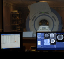
GE Signa HDX 3.0T
Bruker 7.0T Animal Preclinical MRI System
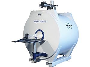
BioSpec 70/30 USR properties:
- Magnetic field strength of 7 T
- 30 cm bore diameter
- 4 parallel receivers
- High performance actively shielded gradient system with integrated shims
- Numerous ready to use MRI methods and sequences available
- State of the art software interface and tools
- Easy routine operation
- Powerful research environment
MR software
ParaVision® 6.0 software package provides a framework for multi-dimensional MRI/MRS data acquisition, reconstruction, analysis and visualization.
This basic software package is supplemented by several extension packages which offer additional experimental techniques or sophisticated post-processing algorithms.
- Standard acquisition, reconstruction, data analysis and visualization
- State-of-the-art 2D and 3D MR imaging techniques
- Easy set-up with auto-adjustments and localizer scans
- Predefined application oriented protocols
- Real-time display of acquired and reconstructed data
- Ready-to-use protocols for ultra-fast imaging sequences including echo-planar imaging (EPI) techniques, fMRI, perfusion imaging, UTE, ZTE, DTI, SWI, Spiral imaging, Cardiac imaging
- On-line, off-line, reconstruction, including baseline correction, window multiplication and phase correction capabilities
MRI Coils
- Single tuned 1H circularly polarized, transmit/receive volume RF coil
- Outer/inner diameter: 198mm/154mm
- Single tuned 1H circularly polarized, transmit/receive volume RF coil
- Outer/inner diameter: 112mm/72mm
- Single tuned 1H circularly polarized, transmit/receive volume RF coil
- Outer/inner diameter: 75mm/40mm
- Single tuned 1H circularly polarized, transmit only volume RF coil
- Outer/inner diameter: 112mm/86mm
- Single tuned 1H 2x2 4 element mouse brain array surface receive coil
- Single tuned 1H 2x2 4 element rat brain array surface receive coil
- Single tuned 10mm ID 1H only surface coil with flexible wire mounting
- Single tuned 20mm ID 1H only surface coil with flexible wire mounting
- Single tuned 30mm ID 1H only surface coil with flexible wire mounting
- Double tuned 1H/31P circularly polarized, transmit/receive volume RF coil
- Outer/inner diameter: 75mm/40mm
- Double tuned 30 mm 1H/31P linearly polarized, receive surface RF coil
Two gradient sets:
- 20 cm ID, 300 mT/m gradient strength, 1080 T/m/s max linear slew rate
- 11.4 cm ID, 660 mT/m gradient strength, 4570 T/m/s max linear slew rate
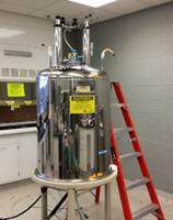
Bruker 9.4T
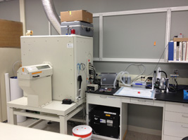
eXplore Locus Micro CT Scanner
Technology Type: volumetric conebeam CT
Analysis Software: eXplore MicroView
Version: 2.2
Features:
2D & 3D image viewing
- Multiple format import & export
- Volume reorientation & alignment
- Histogram plotting
- Analysis data export
- Volume rendering
- Line profiling & measurement
- Quantitative Analysis (BMD, BVF)
- Stereological Parameters (trabecular number, spacing, thickness, connectivity, isotropy)
X-ray source: Fixed tungsten anode
Maximum tube potential: 80 kVp
Maximum tube current: 0.5 mA
Maximum transverse FOV: 40 mm
Maximum radiation exposure level 5 cm from any accessible exterior surface: less than or equal to 0.5 mR/h
Detector design: scintillator and CCD camera, connected with fiber optics
Detector type: Camelia M1 CL 8M
Frame grabber type: Coreco Sapera
Frame grabber model: X64-CL Express 1
Scanner type: RS80
Scanner subtype: 2
Tube type: SRI